Wednesday, February 18, 2009

UV Light-enhanced Tooth Bleaching Dangerous To Eyes And Skin

The light treatment gives absolutely no benefit over bleaching without UV, and damages skin and eyes up to four times as much as sunbathing, reports a study in Photochemical & Photobiological Sciences.

The treatment is at least as damaging to skin and eyes as sunbathing in Hyde Park for a midsummer’s afternoon – one lamp actually gave four times that level of radiation exposure.

And as with sunbathing, fair-skinned or light-sensitive people are at even greater risk, said lead author Ellen Bruzell of the Nordic Institute of Dental Materials.

Bruzell also found that bleaching damaged teeth. She saw more exposed grooves on the enamel surface of bleached teeth than on unbleached teeth. These grooves make the teeth more vulnerable to mechanical stress.

Tooth bleaching is one of the most popular cosmetic dental treatments available. It uses a bleaching agent – usually hydrogen peroxide – to remove stains such as those from red wine, tea and coffee, and smoking.

UV light is claimed to further activate the oxidation process, improving bleaching efficiency. The authors of this Photochemical & Photobiological Sciences article say there is very little substantive evidence to support this claim, and their new study finds no benefit to using UV light.

New Tooth Cavity Protection: Nanoparticles Make Surface Too Slippery For Bacteria To Adhere


Clarkson University Center for Advanced Materials Processing Professor Igor Sokolov and graduate student Ravi M. Gaikwad have discovered a new method of protecting teeth from cavities by ultrafine polishing with silica nanoparticles.

The researchers adopted polishing technology used in the semiconductor industry (chemical mechanical planarization) to polish the surface of human teeth down to nanoscale roughness. Roughness left on the tooth after the polishing is just a few nanometers, which is one-billionth of a meter or about 100,000 times smaller than a grain of sand.

Sokolov and Gaikwad showed that teeth polished in this way become too “slippery” for the "bad" bacteria that is responsible for the destruction of dental enamel. As a result the bacteria can be removed fairly easily before they cause damage to the enamel.

Although silica particles have been used before for tooth polishing, polishing with nanosized particles has not been reported. The researchers hypothesized that such polishing may protect tooth surfaces against the damage caused by cariogenic bacteria, because the bacteria can be removed easily from such polished surfaces.

Monday, February 16, 2009

Eye and Teeth Development Linked to Same Protein

Scientists from the University of Leeds have found a link between eye and teeth formation in the protein CNNM4.
The findings will be reported in American Journal of Human Genetics.

In the Gaza Strip, scientists studied residents who suffer from a genetic condition that causes blindness early in life, as well as significant tooth decay. The condition has been named Jalili syndrome after one of the research team members, Ismail Jalili. A protein known as CNNM4 can be found in the cells responsible for laying down tooth enamel and retinal layers. It may also carry calcium and magnesium to other cells. A mutation in CNNM4 is responsible for Jalili syndrome, which can be transferred genetically to children.

Now that scientists know that the protein is responsible for tooth and eye development, they hope to research gene therapy to battle the condition. Finding this link may also lead to progress in understanding inherited blindness.

ViziLite Plus......for Qral Qancer screening

Using ViziLite Plus along with a standard oral cancer examination improves our ability to identify suspicious areas at their earliest stages. ViziLite Plus is similar to proven early detection procedures for other cancers such as mammography, Pap smear and PSA. ViziLite Plus is a simple and painless examination that gives the best chance to find oral abnormalities at the earliest stage. Early detection of pre-cancerous tissue can minimize or eliminate the potentially disfiguring effects of oral cancer and possibly save your life.

The ViziLite Plus Screening Process:

After a conventional oral cancer examination:

1. The patient rinses with ViziLite Solution for 30 – 60 seconds.

2. The oral cavity is illuminated withthe ViziLite light wand.

3. The dentist looks for and documents any abnormalities.

Healthy tissue absorbs ViziLite illumination. Abnormal tissue reflects it and appears white.

Appearance of abnormal tissue with and without ViziLite illumination.

4. If an abnormality is found a toludene blue solution (Tblue630) is swabbed onto it and photographs are taken for inclusion in the patient record and follow-up.

Appearance of abnormal tissue without and with Tblue630 staining

Saturday, February 14, 2009

Simroid


We've finally created the robot version of Hell: force one to sit, eternally, in a dentist chair and have it's mouth operated on. Could we make it worse? Why not give the bot sensitive teeth, eh? Unfortunately for the Simroid, Kokoro went the whole nine yards when the company designed it to act as a responsive training aid for dentists — teeth and all.

Simroid is designed to simulate a human patient. It can follow spoken orders and react to what's happening. Sensitive teeth allow the robot to know where the trainee is poking around and if said trainee blunders. Simroid responds in appropriate discomfort, visibly grimacing, moving its arms, and telling the doc-hopeful that it hurts. It also has silicon skin that's soft to the touch and air-powered muscles enable its realistic gestures. The designers found that a Simroid garners a better response from dentist than an inert dummy.

Kokoro, a division of Sanrio (known for making Hello Kitty products), showed the Simroid at the 2007 International Robot Exhibition in Tokyo,

To watch video : http://www.youtube.com/watch?v=Vaf-QxhQh6g

DIASTEMA CLOSURE : A CASE

Pre-operative frontal view.
Pre-operative smile.
Pre-operative Left lateral view.
Pre-operative right lateral view
Etching of tooth with adjacent area protected
Characterisation of enamel layer with white tint
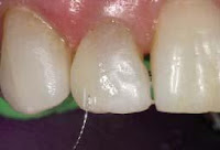
Finishing with aluminium oxide discs.
finishing the restorations.
Egg shaped carbide bur used in the lingual concavities
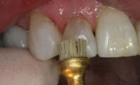
Silicon carbide brush.
Frontal view of anterior teeth
Right lateral view.
Left lateral view.

DIASTEMA CLOSURE 3

Final shaping and polishing is achieved with burs, sandpaper disks, rubber wheels, points, cups, and polishing pastes. Mesial distal dimension is measured on the restored tooth and compared to the distal mesial dimension of the adjacent tooth and space. Adjustments are made to the restored tooth with burs or sandpaper disks. Restoration of the adjacent tooth is achieved using the same technique. Close approximation of composite to composite on the adjacent tooth is achieved by holding the matrix against the adjacent composite with an instrument and light curing.

DIASTEMA CLOSURE 2

The back of the mouth is a dark area because it receives no light. Composite must block out darkness or a restoration appears dark. Placement of opaque material to the lingual covered with translucent material to the facial achieves a natural looking restoration that is not influenced by this darkness. Blending composite color to tooth color is further achieved by proper composite selection, placement and preparation design.
Restoring small diastemas or restoration of teeth that have a large buccal lingual dimension do not require placement of lingual opaque composite.Bonding to enamel provides strength to hold composite onto tooth structure and minimize microleakage. Removal of caries often creates areas of mechanical locking that aids retention. Strength of enamel bonding is increased by beveling across enamel rods. A longer bevel or chamfer preparation creates more surface area f
or strength and provides a long gradual show through of tooth color for better color transition. A translucent outer layer of composite provides a chameleon effect picking up and showing through surrounding color.
Gingival control eliminates a black triangle in the papillae area. Placement of composite subgingival is achieved by placement of a matrix that reflects gum tissue to allow bonding and composite placement.

Tooth structure is prepared, a plastic matrix placed, etching and bonding completed on one tooth.
A lingual wall of composite placed trying to achieve ideal interproximal contours and light cured. Dimensions are made exact or too large. Cured composite is difficult to add to when the oxygen inhibited layer is lost but it is easily removed.
Wrapping a matrix is avo
ided because it produces a straight contour and eliminates the oxygen inhibited layer. A contoured matrix or hand shaping produces convex interproximal areas. A layer of translucent composite is placed across the facial aspect, shaped with hand instruments and light cured.

Friday, February 13, 2009

DIASTEMA CLOSURE

A diastema is a space between front teeth. Diastemas are closed by orthodontics or restoration. A highly successful technique is addition of composite. A space which is too large and closed with composite results in teeth that are esthetically too wide and orthodontics is recommended. Space closure requires placement of composite two adjacent teeth.
Placement of composite onto one tooth can be done it proper tooth dimensions allow it.
Anesthetic is not required unless dentin or root structure is involved. Diamond burs prepare tooth structure creating a rough surface for improved bond strength and to produce bevels that show through tooth color at restoration cavosurface areas. Cross section of enamel rods improves enamel bond strength.
Free ads

Free AdvertisingCoupon CodeDell CouponGap CouponTarget Coupon